Exploring Chest X-Ray Applications and Interpretations

Article Overview
Purpose of the Article
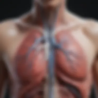
This article offers a detailed examination of chest X-rays, often abbreviated as CXRs, including their significance in the medical landscape. Aiming to bridge theory with practical application, we delve into how CXRs aid in the diagnosis and management of various health conditions. By exploring the technical aspects behind radiographic imaging, we aim to illuminate how CXRs have evolved and why they remain a cornerstone in modern diagnostics.
Relevance to Multiple Disciplines
The implications of chest X-rays extend beyond the realm of traditional medicine. They are integral to disciplines like emergency medicine, pulmonology, and even veterinary practices. Understanding the art and science behind CXR can provide professionals in various fields with the skills needed for accurate diagnoses and effective patient care.
Research Background
Historical Context
The journey of chest X-rays began in the late 19th century with Wilhelm Conrad Röntgen’s groundbreaking discovery of X-rays in 1895. This revolutionary advancement laid the groundwork for current imaging techniques, allowing for non-invasive visualization of internal body structures. Initially used for foreign body detection, CXRs quickly adapted to include applications for diagnosing pneumonia, tuberculosis, and heart conditions. Over decades, continuous innovations, particularly in digital imaging, have significantly enhanced the efficiency and clarity of CXR results.
Key Concepts and Definitions
To fully appreciate the nuances of chest X-rays, it's essential to comprehend a few fundamental concepts.
- Radiographic Imaging: The process of capturing images of the internal structure using X-ray technology.
- Contrast: Refers to the difference in image density, which aids in revealing detailed anatomical features.
- Interpretation: The act of analyzing X-ray images to identify pathological conditions, requiring a skilled eye and solid foundational knowledge.
"Radiographs are like a puzzle; it takes intuition and expertise to piece together the clues they provide about a patient's condition."
"Radiographs are like a puzzle; it takes intuition and expertise to piece together the clues they provide about a patient's condition."
Understanding these concepts is foundational for those in the medical field who rely on CXRs for effective diagnosis and treatment plans. The following sections will expand on their applications, technological advancements, and interpretation of results, ensuring a robust comprehension of CXRs in clinical practice.
Intro to CXR X-Ray
Chest X-rays (CXRs) have long stood as one of the cornerstones in medical imaging, serving not only as a diagnostic tool but also as an indispensable aid in evaluating a range of thoracic conditions. The importance of this topic lies in understanding both the essential role CXRs play in contemporary medicine and the methodologies that underlie their use. By grasping the intricacies of CXR, professionals in the field can better appreciate how these images inform clinical decisions and enhance patient care.
At a basic level, CXRs are essential for evaluating the respiratory system and the structures nearby. They're primarily used to investigate ailments such as pneumonia or congestive heart failure, where swift and accurate imaging is crucial. In an age where healthcare strives for efficiency and accuracy, CXRs effectively provide a non-invasive glimpse into the inner workings of the chest, guiding physicians in making informed decisions about further diagnostic tests or treatment options.
Benefits of Understanding CXRs
- Immediate Visual Insight: CXR gives quick answers, making it a first-line tool in emergency settings.
- Wide Accessibility: Most healthcare facilities have the equipment necessary to perform chest X-rays, allowing for broad utility across different healthcare settings.
- Cost-Effectiveness: Compared to advanced imaging technologies like CT scans, CXRs are typically less expensive, yet they provide substantial diagnostic value.
When discussing CXRs, consideration must also be given to the potential for misinterpretation. Images might appear deceptively clear, masking underlying conditions that require further investigation. Hence, the importance of honing interpretive skills cannot be overstated; detailed knowledge about what to look for can mean the difference between a missed diagnosis and timely intervention.
As research progresses, the advent of digital technologies and AI applications also promises to enhance our understanding and interpretation of CXR images, ensuring this modality remains relevant in modern diagnostics. Thus, understanding CXRs isn't just about recognizing their historical significance or mastering techniques; it's about embracing the evolution of their applications in the continually changing sphere of healthcare.
"Chest X-rays are like a window to the thorax—providing vital insights, yet requiring a careful eye to interpret fully.”
"Chest X-rays are like a window to the thorax—providing vital insights, yet requiring a careful eye to interpret fully.”
In the next sections, we will delve deeper into the definition and purpose of CXRs, along with their fascinating historical context.
Technical Foundations of CXR X-Rays
Understanding the technical foundations of CXR X-rays is pivotal for grasping their significance in the medical field. These foundational aspects establish the context in which chest X-rays are utilized, ensuring accurate interpretations and enhancing clinical decision-making. The principles of radiography underpin this area, focusing on how X-ray technology works, the various techniques employed, and the implications of each method.
Principles of Radiography
Radiography hinges on the interaction between X-rays and matter, primarily the different tissues in the human body. The basic principle involves the emission of X-rays from a source, their penetration through the body, and the subsequent capture of the transmitted rays on a detector or film. X-rays, being high-energy photons, have varying levels of absorption depending on the density and composition of the tissues they encounter. This differential absorption is what creates the contrast seen in an X-ray image.
Furthermore, the geometry of the setup plays a crucial role. For chest X-rays, the typical positioning involves the patient standing or sitting, which helps in providing a clear view of the thoracic cavity. A striking feature is the importance of proper alignment to minimize distortion of the anatomical structures, thus allowing for accurate diagnosis.
As technology progresses, digital radiography has emerged as a prominent method, surpassing traditional film-based techniques. It boasts enhanced image quality, reduced radiation exposure, and instant image retrieval, making it a preferred choice in many clinical settings.
Types of Chest X-Ray Techniques
Different techniques employed in chest radiography serve specific purposes and provide distinct views, aiding in the comprehensive evaluation of thoracic structures. Here’s a closer look at each method:
PA View
The posteroanterior (PA) view entails the patient facing the X-ray plate, which captures the thorax's front-to-back projection. A key characteristic of the PA view is that it enables a clear view of heart size and lung fields without significant magnification of the heart. This makes it a popular choice for routine chest X-rays, particularly in assessing conditions like pneumonia and cardiac anomalies. One unique feature is its ability to provide a standardized reference, assisting in easier comparisons over time.
While beneficial for clarity, one must recognize its limitations. The PA view may not reveal behind-obscured structures, leading to potential diagnostic oversights. Thus, further imaging may be necessary for comprehensive evaluations.
AP View
The anteroposterior (AP) view, in contrast, shows the chest from the front to the back, typically used when the patient cannot stand properly, such as in bedridden scenarios. A significant aspect of the AP view is that it can be obtained quickly, making it advantageous in emergency settings. However, a critical downside is that it may result in heart enlargement due to increased magnification from the closer positioning of the X-ray tube. This discrepancy can complicate interpretations, demanding careful consideration of the clinical context.
Lateral View
The lateral view adds depth to the diagnosis by providing a side perspective of the thorax. This view is crucial for evaluating the diaphragm, pleural spaces, and mediastinal structures that might be obscured in other methods. A unique feature of the lateral view is its capability to show the anatomy of the lungs in relation to surrounding organs, thus aiding in identifying pleural effusions that might not be evident in PA or AP views. Nonetheless, it’s worth noting that the lateral view may sometimes be challenging to interpret due to superimposed structures, which can obscure pathology.
Oblique Views
Oblique views involve angling the X-rays at various intervals, typically for the purpose of enhancing visualization of specific structures or lesions. They are particularly useful for examining potential mass lesions or other abnormalities in the lungs and heart that wouldn’t display clearly on standard views. The flexibility of oblique positioning allows radiologists to adjust to patient needs and anatomical variations. However, the interpretation can be complex, as these views might present anatomical distortions that complicate the analysis.
"A strong foundation in principles and techniques elevates the efficacy of CXR interpretations and impacts patient care substantially."
"A strong foundation in principles and techniques elevates the efficacy of CXR interpretations and impacts patient care substantially."
In summary, understanding these technical foundations is not just about grasping the methods of imaging. It’s about appreciating how each view interacts with the clinical context to offer a clearer diagnosis and better patient outcomes.
Clinical Applications of Chest X-Rays
The realm of clinical medicine greatly benefits from the application of Chest X-Rays (CXR). They provide a crucial window into the internal workings of the human body, specifically featuring the lungs and heart. Their ability to quickly highlight abnormalities sets them apart in diagnostic imaging. Clinical applications of CXR not only assist in diagnosing various conditions but also guide treatment decisions in real time, which can be life-saving in emergencies. Moreover, CXR serves as a fundamental assessment tool in both primary and advanced healthcare settings. It is widely regarded as one of the first imaging modalities employed for patients exhibiting respiratory symptoms, underlining its relevance.
Diagnosis of Respiratory Conditions
Pneumonia
Pneumonia is an inflammation in the lungs, often caused by infections. Among its diagnostic tools, CXR stands out for its speed and efficiency. An X-Ray can reveal characteristic signs of pneumonia, such as segmental or lobar consolidation. These specific findings allow healthcare professionals to discriminate between types of pneumonia and subsequently tailor treatment accordingly. A unique aspect of pneumonia illustrated on a CXR is the presence of air bronchograms, which can serve as an indicative marker. But, even as CXRs are a first-line imaging choice for pneumonia diagnosis, they might not detect early-stage infections, thereby necessitating further evaluation in some cases.
Tuberculosis
Another major respiratory condition that benefits from CXR is tuberculosis (TB). This communicable disease can be insidious in nature, often laying dormant for years. However, CXR can unearth typical features such as cavitary lesions or nodules in the lungs that are indicative of TB reactivation. TB's hallmark of upper lobe infiltrates is easily identifiable on X-Rays, making it a critical choice for detecting active infections. Despite its effectiveness, X-Rays are not infallible; false negatives can occur, especially in immunocompromised patients or early stages of the disease, highlighting a potential drawback to its sole use in diagnostics.
Lung Cancer
Lung cancer, a prevalent and deadly disease, presents another critical application of CXR. X-Rays may reveal masses or abnormalities that could indicate malignancy. They often serve as a preliminary screening tool, providing critical visual clues that prompt further investigation, such as a CT scan or biopsy. What makes lung cancer particularly challenging is the variance in X-Ray appearances; nodules can range in size and shape, making it vital for trained professionals to differentiate between benign and malignant entities. However, following a negative initial CXR, there remains a risk of symptoms being ignored, which might delay diagnosis and treatment.
Evaluating Cardiac Abnormalities
Cardiomegaly
CXR is invaluable in evaluating heart conditions, most notably in identifying cardiomegaly, an enlargement of the heart. This condition may be indicative of various underlying issues such as hypertension or heart failure. On an X-Ray, an enlarged cardiac silhouette can signal the need for immediate further investigation. Nonetheless, the interpretation of cardiomegaly can be subjective, as variations in individual anatomy and positioning may affect the results. Despite these challenges, its identification is integral to appropriate patient management.
Heart Failure
Heart failure, characterized by the heart's inability to pump sufficiently, can also be assessed through CXR. The classic X-Ray findings, such as pulmonary congestion or pleural effusion, help clinicians gauge the severity of the condition quickly. This rapid assessment can guide immediate treatment strategies and enhance patient outcomes. However, just like cardiomegaly, CXR has its limitations, as it cannot determine the underlying cause of heart failure on its own, necessitating additional tests for a comprehensive evaluation.
Pericardial Effusion
Pericardial effusion, an accumulation of fluid in the pericardial space surrounding the heart, is another condition where CXR proves crucial. The X-Ray can reveal signs of an enlarged cardiac silhouette indicating fluid presence, which is essential for timely intervention. The particular challenge with pericardial effusion lies in its variable presentation; small effusions may go undetected on an X-Ray. Thus, while it plays a significant role in early screening, additional imaging methods like ultrasound may be warranted for confirmation.
Assessment of Trauma
Fractures
In trauma cases, CXR has become a go-to for evaluating potential rib or thoracic spine fractures. It efficiently highlights fractures or dislocations, guiding urgent surgical interventions when needed. The utility of CXR in this context is unmatched due to its rapid nature, allowing for quicker treatment decisions. However, one must be cautious; while noteworthy rib fractures may show up on X-Rays, minor or non-displaced fractures can be elusive, which may lead to misdiagnosis.
Pulmonary Contusions
Pulmonary contusions are another injury type that can be assessed using CXR. These bruises of lung tissue typically occur after blunt chest trauma. X-Rays can reveal opacities, indicative of pulmonary contusions, which may not be immediately apparent. These findings are critical for forming a treatment plan in trauma patients. However, the initial CXR may not capture all the damage done, so monitoring via follow-up imaging is often essential to ensure effective management.
Interpreting Chest X-Rays


Interpreting chest X-rays (CXR) is not merely an art but a crucial skill in the realm of medical diagnostics. Understanding the nuances involved in reading these X-rays can drastically affect patient care and clinical decision-making. The ability to correctly interpret CXRs allows healthcare providers to identify various pathologies, monitor treatment responses, and guide further investigative processes. Without a solid grasp on interpretation, one might easily overlook significant details that could aid in patient assessments.
Basic Principles of Interpretation
At its core, interpreting a CXR demands a systematic approach. Radiologists are trained to examine images using a set of guiding principles, ensuring they don’t miss essential findings. Here’s a basic framework:
- Review the patient’s clinical history: Always correlate findings with the patient’s symptoms and history.
- Assess image quality: Check for factors like rotation, inspiration level, and exposure as they heavily influence interpretations.
- Use a systematic approach: Typically, radiologists will evaluate the images from top to bottom, looking at the lungs, heart, vascular structures, bony thorax, and diaphragm.
- Look for patterns: Familiarity with common patterns helps in forming an initial impression that can guide further investigation.
Developing this methodology is crucial as it helps mitigate the risk of missing subtle abnormalities.
Common Findings and Patterns
Consolidation
Consolidation refers to a process where lung tissue becomes firm and solid due to various conditions, primarily infection. This aspect is critical as it signals that the alveoli are filled with fluid, pus, or cells instead of air, often seen in conditions like pneumonia.
- Key Characteristics: On a CXR, this may appear as a dense, localized area changing the typical lung silhouette.
- Benefits: Understanding consolidation can lead to quicker diagnoses and treatment interventions, making it a cornerstone of CXR interpretation.
- Advantages/Disadvantages: However, its presence on an X-ray doesn’t always clarify the underlying cause, necessitating further tests.
Pleural Effusion
Pleural effusion involves the buildup of fluid in the pleural space. Interpreting this correctly is vital, as it may indicate a range of serious conditions.
- Key Characteristics: It often presents as a meniscus sign on the X-ray, which is visualized as a distinct curve of fluid along the lung margin.
- Benefit: Early identification can prompt immediate management, which is crucial in cases such as heart failure or malignancy.
- Unique Feature: The challenge with pleural effusions is determining whether it's transudative or exudative in nature, influencing treatment pathways.
Hyperinflation
Hyperinflation is an increase in air within the lungs due to obstructive pulmonary diseases like Chronic Obstructive Pulmonary Disease (COPD). Identifying this correctly can guide therapeutic approaches and lifestyle modifications.
- Key Characteristics: On the CXR, you may observe flattened diaphragms and increased retrosternal airspace.
- Benefits: Recognizing hyperinflation is critical as it often correlates with patients requiring further pulmonary rehabilitation, chronic management, or possible surgical interventions.
- Advantages/Disadvantages: While visible on X-ray, its assessment can sometimes be misleading without clinical context, as not all patients with hyperinflation exhibit symptoms.
Errors and Pitfalls in Interpretation
Despite established principles and common findings, errors in interpretation can and do occur. A few pitfalls include:
- Failing to correlate symptoms with findings, leading to missed diagnoses.
- Misinterpreting overlapping structures, especially when images aren’t correctly aligned.
- Overlooking subtle signs of disease due to incomplete or hurried reviews.
Overall, increasing familiarity with common conditions and refining interpretative skills through continuous education is paramount. Accumulating experiences from varied cases can enhance confidence and accuracy in interpreting chest X-rays.
Advancements in Chest X-Ray Technology
The evolution of chest X-ray (CXR) technology marks a pivotal turning point in the field of medical imaging. As the medical landscape continually shifts, embracing new methodologies becomes imperative. These advancements not only enhance diagnostic accuracy but also improve patient safety and workflow efficiency. The integration of novel techniques into CXR practices reshapes how healthcare professionals diagnose and treat various conditions.
Digital Radiography
Digital radiography is considered a transformative leap from traditional film-based imaging. With its capability to convert X-ray data into digital images, this technology provides several distinct advantages. Speed is a notable benefit; images can be ready in mere seconds, significantly cutting down patient wait times. Moreover, the quality of images improves markedly, offering enhanced contrast and resolution.
In terms of storage and sharing, digital X-rays are superior. Unlike physical films, which can deteriorate or be easily misplaced, digital files can be stored in vast databases and shared rapidly across departments or institutions. Clinicians, therefore, can access patient records with just a few clicks, streamlining collaboration.
Despite its benefits, digital radiography isn't without its challenges. The initial setup cost can be quite high, which may make the transition daunting for some healthcare facilities. Plus, training staff to fully harness the potential of these systems is also essential, ensuring they both understand the technology and maintain proficiency.
Artificial Intelligence in Imaging
The incorporation of artificial intelligence (AI) into chest X-ray interpretations is revolutionizing diagnostic accuracy. Algorithms powered by machine learning can analyze images with remarkable precision. Healthcare professionals often find themselves collaborating with these smart systems, which can detect anomalies that might escape the naked eye.
To illustrate, AI tools can flag potential conditions like pneumonia or signs of lung cancer almost instantaneously. This aids radiologists in prioritizing cases and making informed decisions quickly. The overall effectiveness increases as healthcare providers can provide timely treatments, thereby improving patient outcomes.
However, the implementation of AI must be approached with caution. Concerns regarding over-reliance on AI systems exist. Human oversight remains crucial, as technology may sometimes stumble or misinterpret data. Maintaining a balance where AI serves as a complementary tool helps mitigate these risks, allowing clinicians to engage in informed analysis.
Tele-radiology Applications
Tele-radiology is a crucial development that has surged in use, particularly during the recent global health crises. This technology represents a paradigm shift in how radiographic services are delivered. Physicians can remotely access and interpret CXR images, breaking down geographical barriers in patient care.
One of the primary advantages of tele-radiology is its ability to facilitate swift consultations. For instance, if a patient in a rural area requires a chest X-ray, the images can be sent instantly to a specialist in an urban center. This capability can lead to quicker diagnosis and treatment initiation, thus saving lives.
Moreover, tele-radiology also supports continuous learning without geographical constraints. Radiologists can confer with peers from across the globe, exchanging knowledge and insights on complex cases, enhancing professional development. Nevertheless, as is the case with many technologies, there are security and privacy concerns associated with transmitting sensitive patient data online. Thus, stringent measures must be in place to protect patient confidentiality.


In summary, advancements in chest X-ray technology are not merely enhancements but essential developments that improve diagnostics, patient care, and collaborative ability.
In summary, advancements in chest X-ray technology are not merely enhancements but essential developments that improve diagnostics, patient care, and collaborative ability.
Safety Considerations in CXR Imaging
In the realm of medical imaging, safety considerations have become paramount, especially when it comes to chest X-rays (CXR). The practice of radiography, though invaluable, comes with its set of concerns pertaining to radiation exposure. Addressing these issues not only underscores the responsibilities that radiologists must uphold but also enhances trust between healthcare providers and patients. A thoughtful approach to safety can yield numerous benefits, thereby lifting the field to new heights of proficiency.
Radiation Exposure and Risks
The benefits of CXR in diagnosing various conditions cannot be overlooked. However, it is crucial to recognize that X-rays utilize ionizing radiation, which, if mismanaged, can lead to potential risks for the patient. A fundamental aspect of medical ethics revolves around minimizing harm; hence, understanding the intricacies of radiation exposure is essential.
- Nature of Risk: The primary concern involves the cumulative effect of radiation exposure over time. Prolonged or unnecessary exposure can result in an increased risk of cancer, particularly in sensitive groups like children, who are more susceptible to radiation.
- Dose Management: It’s also essential to recognize that not all X-rays are created equal. The dose levels vary significantly depending on the technique and technologies in use. For instance, a computed tomography (CT) scan delivers a higher dose compared to a standard chest X-ray, raising concerns regarding appropriateness in imaging choices for specific conditions.
"The key to navigating the landscape of CXR safely lies in both understanding the risks and implementing diligent practices to mitigate them."
"The key to navigating the landscape of CXR safely lies in both understanding the risks and implementing diligent practices to mitigate them."
Best Practices for Minimizing Risk
To tackle the concerns raised by radiation exposure, several best practices can be adopted to provide a framework for safe and effective CXR utilization.
- Appropriateness of Imaging: Before proceeding with a CXR, a rigorous review of the patient's history and clinical indicators should be conducted. Imaging should only be performed when it is warranted, ensuring that the benefits outweigh the risks.
- Use of Correct Protocols: Healthcare practitioners should utilize established protocols that are focused on minimizing radiation dose while ensuring image quality. Techniques such as:
- Shielding and Protective Gear: Implementing lead aprons and thyroid shields for patients can significantly limit unnecessary dose exposure. It's also wise to shield adjacent organs that aren’t the focus of the imaging.
- Education and Awareness: Continuous training for healthcare professionals on the importance of radiation safety can lead to better decision-making. Keeping abreast of the latest research and guidelines contributes to a culture of safety in medical imaging.
- Patient Communication: Clear communication with patients regarding the risks and benefits of their procedures can foster an environment of trust. Patients should feel empowered to ask questions and express concerns about their imaging.
- Evaluate the necessity for X-ray—consider if alternative modalities, like ultrasound, could serve the diagnosis more safely.
- Proper patient positioning to avoid repeat exposures
- Adjusting the X-ray machine settings according to the patient's age and body type
Future Directions in CXR Research
As we forge ahead in the realm of medical imaging, understanding the future directions in CXR research becomes increasingly crucial. With the rapid advancements in technology and a growing demand for efficiency in diagnostic processes, there’s an undeniable need to explore how Chest X-rays could evolve to meet future challenges. The emphasis on improved accuracy and reduced healthcare costs is at the forefront of this exploration.
Emerging Imaging Techniques
The landscape of imaging techniques for CXRs is set to expand dramatically in the coming years. Developers and researchers are currently focused on innovating methodologies that enhance the clarity and diagnostic capability of these essential tools. For instance, promising techniques such as spectral imaging are becoming more prevalent. Unlike conventional X-rays which rely on a single intensity measurement, spectral imaging captures multiple energy levels, granting radiologists a richer perspective of anatomical structures and pathological changes.
This technique enables professionals not only to detect diseases earlier but also to assess them more accurately. Another avenue of exploration is the integration of 3D chest imaging, which allows for a detailed visualization of complex anatomical relationships that traditional two-dimensional CXRs cannot provide. By investing in such emerging technologies, the goal is to elevate the standard of care, especially in high-stakes clinical settings.
Furthermore, Advanced software algorithms using machine learning techniques are showcasing an ability not previously seen. These can analyze vast datasets quickly, pinpointing abnormalities with impressive precision. The future may well hold CT-like quality in diagnostics derived from simple CXR techniques due to these advancements, which could make them a go-to option for primary health settings as well.
Integration with Other Modalities
Another significant future direction is the seamless integration of CXRs with other imaging modalities such as computed tomography (CT) and magnetic resonance imaging (MRI). This multidisciplinary approach could yield comprehensive diagnostic insights that aren’t possible in isolation.
By synthesizing data from CXRS with CT’s intricate images, clinicians have a better outlook on conditions like lung cancer, where precise staging is critical. Moreover, tools that combine functional imaging with anatomical imaging will positively impact treatment planning and follow-ups, enabling multidisciplinary teams to tailor interventions much more effectively.
The cooperative use of images generated from varied procedures could, for example, lead to improved outcomes in trauma care, as it allows medical teams to have a holistic view of the patient.
"The future of CXRs is promising, as their potential expands beyond mere diagnostic tools, becoming integral components in the holistic view of patient care."
"The future of CXRs is promising, as their potential expands beyond mere diagnostic tools, becoming integral components in the holistic view of patient care."
As we venture into these collaborative relationships between imaging types, we bolster our ability to make diagnoses that are not only timely but also nuanced, which ultimately enhances patient safety and outcomes. It’s an exciting time in the realm of medical imaging, where CXRs, armed with evolving technologies and integrative strategies, will undoubtedly hold their ground as a pivotal player in diagnostics.
Culmination
The conclusion serves as the cornerstone of the discussion on chest X-rays, notably stitching together the various threads presented in the article. Here, we underline the importance of recognizing CXRs not just as mere imaging tools but as essential instruments that facilitate accurate diagnostics and treatment plans. By reflecting on the aptitude of chest X-rays in unveiling a multitude of respiratory and cardiac disorders, we can appreciate their pivotal role in patient management.
Summarizing Key Insights
In summary, the examination of CXRs reveals several key takeaways:
- Versatility: Chest X-rays are valuable for diagnosing a broad spectrum of conditions, ranging from pneumonia to cardiomegaly.
- Technological advancements: The evolution from traditional techniques to digital radiography and AI integration showcases the continuous improvement in imaging practices.
- Safety measures: Emphasizing the importance of minimal radiation exposure is crucial for protecting both patients and healthcare workers.
- Interpretation skills: Accurate readings hinge on recognizing common patterns and avoiding pitfalls, which underscores the importance of ongoing education in radiology.
Conclusively, the integration of theoretical knowledge with practical interpretation fosters better patient outcomes.
The Ongoing Role of CXR in Medicine
As we look ahead, it is clear that chest X-rays will continue to play a vital role in the medical landscape. They are not just diagnostic tools; they represent a bridge between clinical suspicion and concrete findings. CXRs will adapt to augment their capabilities through enhanced imaging techniques and digital platforms. The use of artificial intelligence and tele-radiology, for instance, is setting the stage for heightened efficiency in reading X-rays and sharing interpretations across distances, making expert advice readily available even in remote areas.
Moreover, as newer modalities emerge, the challenge remains to integrate these technologies seamlessly while ensuring that chest X-rays retain their irreplaceable value. The knowledge around their interpretation must also evolve, emphasizing interdisciplinary approaches that harness both radiological and clinical expertise.
In essence, the journey into the realm of chest X-rays isn't just a matter of technology and methodology; it is about constantly enhancing patient care through informed and timely medical decisions.


