Innovations in Lung Imaging: Techniques and Trends


Article Overview
Purpose of the Article
This article aims to investigate the multifaceted domain of lung imaging. It will detail various imaging modalities, focusing on their significance in respiratory health. The purpose is twofold: first, to inform readers about the technological advancements that improve diagnosis and treatment of lung conditions; second, to explicate the impact of artificial intelligence in lung imaging interpretation.
Relevance to Multiple Disciplines
Lung imaging is not merely a medical specialty; it intersects various fields such as radiology, pulmonology, and artificial intelligence. Professionals from different disciplines can benefit from understanding how imaging works and its implications for patient care. Educational institutions and healthcare organizations increasingly prioritize this knowledge in curriculum and training programs, indicating its growing importance across multiple domains.
Research Background
Historical Context
The history of lung imaging spans over a century, beginning with the discovery of X-rays. The evolution from basic X-ray imaging to advanced modalities like CT and MRI has fundamentally altered the approach to diagnosing lung diseases. Each technological leap has introduced new capabilities and insights into respiratory health, reshaping clinical practices and public health approaches.
Key Concepts and Definitions
Understanding the methods used in lung imaging requires familiarity with essential terms:
- X-ray: A form of electromagnetic radiation used to view structures within the body. It's fast and widely accessible.
- CT (Computed Tomography): This imaging technique generates detailed images of internal organs and structures, offering enhanced visibility compared to standard X-rays.
- MRI (Magnetic Resonance Imaging): Utilizes magnetic fields and radio waves to create high-resolution images of tissues, providing contrasting capabilities that are particularly useful in soft tissue assessment.
- Ultrasound: An imaging method that uses sound waves to produce images of organs, which is beneficial for guiding procedures or evaluating conditions that require real-time assessment.
"Lung imaging technology is at the forefront of diagnosing conditions at earlier stages, where treatment efficacy is often greatest."
"Lung imaging technology is at the forefront of diagnosing conditions at earlier stages, where treatment efficacy is often greatest."
Beyond these technical definitions, the incorporation of artificial intelligence in imaging is a key concept. AI algorithms assist radiologists by analyzing images to detect abnormalities, enhancing diagnostic speed and accuracy. Its role in future innovations and how it can optimize workflows remains a critical area for exploration throughout this article.
Intro to Lung Imaging
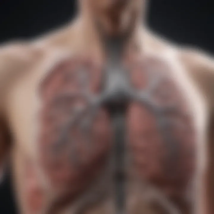
Lung imaging serves as a critical pillar in modern medicine, particularly in the field of respiratory health. Through various imaging modalities, it provides essential insights into the structure and function of the lungs, aiding in the accurate diagnosis and management of pulmonary diseases. Understanding the complexity of lung imaging is crucial given the increasing prevalence of respiratory conditions worldwide. With improved imaging techniques, healthcare professionals can detect abnormalities and diseases at earlier stages, significantly impacting patient outcomes.
The introduction discusses not only the methods employed in lung imaging but also the applications and innovations that continue to evolve in this field. Radiologists, clinicians, and researchers all benefit from an enhanced understanding of lung imaging modalities. This holistic overview serves to bridge the gap between technical detail and clinical practice, making it relevant for professionals dedicated to respiratory health.
Importance of Lung Imaging in Medicine
Lung imaging is indispensable in the medical field for several reasons. First, it enables healthcare providers to visualize and assess the lungs' condition, which is vital for diagnosing various lung diseases, such as pneumonia, lung cancer, and chronic obstructive pulmonary disease (COPD). Second, it assists in monitoring disease progression, treatment efficacy, and post-treatment evaluation, allowing for data-driven clinical decisions.
Key benefits of lung imaging include:
- Rapid Diagnosis: Imaging techniques like chest X-rays and CT scans provide immediate visual information, speeding up the diagnostic process.
- Non-invasive Nature: Many imaging modalities are non-invasive, meaning patients can undergo examinations with minimal risk compared to more invasive procedures.
- Guidance for Treatment Approaches: Radiological assessments guide therapeutic interventions, whether surgical or pharmacological.
In essence, lung imaging not only plays a role in diagnosis but also shapes the entire management of pulmonary conditions, reinforcing the importance of continuous advancement in imaging technology.
Historical Development of Imaging Techniques
The historical journey of lung imaging reveals profound advancements that have transformed the diagnostic landscape. Initially, imaging relied on rudimentary techniques, primarily film-based X-rays, which offered limited insights into lung pathology. Over the decades, technological innovations have led to the development of advanced imaging methods, allowing deeper scrutiny of lung anatomy and function.
Key milestones in the evolution of lung imaging are:
- X-ray Imaging: Discovered by Wilhelm Conrad Röntgen in 1895, X-rays became the first method for visualizing the lungs. Their simplicity and speed revolutionized medical diagnostics.
- Computed Tomography (CT): Introduced in the 1970s, CT scans deliver cross-sectional images, vastly improving the detail of lung assessments.
- Magnetic Resonance Imaging (MRI): Though less common in lung imaging due to respiratory motion, MRI has offered new perspectives on lung structure and function since the 1980s.
- Ultrasound Technology: Emerging over recent decades, ultrasound presents unique opportunities for assessing lung conditions, especially in bedside applications.
Understanding the historical context of lung imaging can illuminate the significance of current methods and encourage the pursuit of future innovations.
Key Imaging Modalities for Lung Assessment


The assessment of lung health and disease relies heavily on various imaging modalities. This section explores the most commonly used techniques, illustrating their significance in clinical practice and research. By understanding the strengths and weaknesses of each approach, medical professionals can make informed decisions about patient care and diagnostic processes.
X-ray Imaging: Basics and Limitations
X-ray imaging is one of the most traditional methods employed in lung assessment. It works by passing a small amount of radiation through the body to create images of the internal structures. X-rays can quickly reveal abnormalities such as pneumonia, tumors, or other lung conditions. They provide a straightforward method for initial evaluation.
However, X-rays come with limitations. The images may lack detail, making it challenging to distinguish between different types of abnormalities. For instance, subtle lesions may go undetected. Additionally, X-rays expose patients to radiation, which raises concerns regarding safety, especially in cases requiring multiple imaging sessions. Therefore, while X-ray imaging serves as a useful first step, it is often supplemented by more advanced techniques for a comprehensive diagnosis.
Computed Tomography (CT) Scans: Advanced Visualization
Computed Tomography, or CT scans, offer a significant advancement over traditional X-rays. This modality utilizes X-ray technology to capture multiple slices of images, which are then processed to create a detailed 3D representation of the lungs. CT scans enhance visualization, allowing for better identification of lung diseases, structural abnormalities, and tumors.
The precision of CT images aids in assessing conditions like chronic obstructive pulmonary disease (COPD) and pulmonary nodules. Furthermore, CT scans can be performed with different protocols, such as high-resolution or contrast-enhanced scans, to tailor the examination to specific clinical questions. However, similar to X-rays, CT scans involve exposure to radiation, necessitating careful consideration of their use, particularly in younger populations or patients requiring repeated assessments.
Magnetic Resonance Imaging (MRI) in Pulmonary Diagnostics
Magnetic Resonance Imaging, or MRI, is less commonly used for lung imaging compared to X-rays or CT scans. This is due to the complex air-tissue interaction within the lungs, which can obscure the clarity of the images. MRI is advantageous in soft tissue visualization and may be employed when evaluating pleural diseases or mediastinal structures.
The benefits of MRI include the absence of ionizing radiation and the ability to capture images in various planes. This modality may provide critical information about lung tumors, especially when characterizing their spread to adjacent structures. However, MRI tends to be less effective in detecting certain lung conditions, and the longer imaging time can be a limitation for some patients.
Ultrasound Imaging: Emerging Applications in Lung Health
Ultrasound imaging has traditionally been underused in lung assessment, but its role is expanding. It uses sound waves to produce images and does not involve radiation, rendering it a safer option for certain patient populations. Ultrasound is particularly useful for assessing pleural effusions, identifying masses, or guiding procedures such as thoracentesis.
Recent advancements have led to increased utilization of ultrasound in emergency settings. It offers real-time imaging, allowing for immediate assessment of conditions like pneumothorax or pulmonary embolism. Despite its advantages, ultrasound has limitations in visualizing deeper lung structures due to air interference. Its effectiveness depends on the operator's skill and the specific situation.
In summary, each imaging modality offers unique strengths and weaknesses. Understanding these can drive clinical decisions, ensuring optimal patient outcomes in lung health assessments.
In summary, each imaging modality offers unique strengths and weaknesses. Understanding these can drive clinical decisions, ensuring optimal patient outcomes in lung health assessments.
Innovations in Lung Imaging Technology
Innovations in lung imaging technology have reshaped the landscape of pulmonary diagnostics. As respiratory health concerns grow, the demand for precise and advanced imaging techniques increases. New technologies bring benefits such as improved accuracy in diagnosis, faster processing times, and the ability to visualize complex lung structures. Understanding these innovations can critically influence treatment plans, enhance patient outcomes, and introduce efficiency in clinical workflows.
Advancements in 3D Imaging Techniques
Three-dimensional imaging techniques represent a significant leap forward in lung diagnostics. Traditional 2D imaging often struggles to convey detailed anatomical structures. In contrast, 3D imaging allows for intricate visualizations of the lung anatomy, including airways, blood vessels, and surrounding tissues.
One of the notable advancements in this area is the utilization of high-resolution computed tomography (HRCT). HRCT provides detailed cross-sectional images, which can be reconstructed into 3D models. These models assist radiologists in identifying subtle pathologies that 2D images may overlook.
Moreover, software solutions that support 3D visualization have become more user-friendly, enabling better collaboration between medical professionals. Clinicians can more effectively discuss cases and tailor treatment plans based on these enhanced visual aids. Improved training tools for medical students and professionals also arise from 3D imaging, allowing them to grasp complex pulmonary anatomy with greater clarity.
Integration of Artificial Intelligence in Image Analysis
The integration of artificial intelligence (AI) in lung imaging marks an era of exponential growth in diagnostics. AI algorithms analyze vast datasets of lung images, recognizing patterns that may be imperceptible to the human eye. This capability enhances diagnostic accuracy, particularly for conditions like lung cancer or interstitial lung diseases.
Machine learning models, for instance, can be trained to differentiate between benign and malignant nodules on CT scans. This empowers radiologists to make more informed decisions. In addition, AI plays a crucial role in automating routine tasks, allowing radiologists to focus on more complex cases.
AI in lung imaging brings not only increased accuracy but also efficiency, revolutionizing pathology detection.
AI in lung imaging brings not only increased accuracy but also efficiency, revolutionizing pathology detection.
Another critical aspect of AI integration is its ability to continuously learn. As more imaging data get fed into these systems, their performance improves over time. Furthermore, AI tools can help standardize imaging protocols and reports, creating consistency across different healthcare facilities.
Clinical Applications of Lung Imaging
Lung imaging plays a vital role in diagnosing and managing various pulmonary conditions. Health professionals rely on these imaging techniques to visualize the lung structure and function, guiding their clinical decisions. This section will discuss the significant applications of lung imaging in clinical practice, highlighting its importance in diagnosing common lung diseases, monitoring lung cancer, and assessing inflammatory and infectious conditions.


Diagnosing Common Lung Diseases
Lung diseases such as chronic obstructive pulmonary disease (COPD), asthma, and pneumonia present varying symptoms that can lead to complex diagnostic challenges. Imaging helps to clarify these conditions by providing detailed visual information.
- X-ray imaging is often the first step in diagnosing lung diseases. It can reveal lung infections, fluid accumulation, and other abnormalities. However, its limitations include a lack of detail in soft tissue differentiation.
- Computed Tomography (CT) scans provide greater clarity, offering cross-sectional views that help identify issues like emphysema, bronchiectasis, and lung nodules. The accuracy of CT scans is crucial for developing targeted treatment plans.
- Magnetic Resonance Imaging (MRI), while less common, can be beneficial in evaluating tumors or lung issues related to vascular structures. It provides superior soft tissue contrast.
Regular advancements in these modalities result in better diagnostic capabilities, ensuring timely and accurate management of lung diseases.
Lung Cancer Detection and Monitoring
Lung cancer is a leading cause of cancer-related deaths worldwide. Early detection is critical for improving prognoses and survival rates. Imaging modalities are key components in lung cancer screening and management.
- Low-dose CT screening has become standard for high-risk populations. By detecting small nodules at an early stage, it allows for earlier interventions.
- Post-diagnosis, imaging plays a crucial role in staging lung cancer. It helps determine tumor size, the extent of metastasis, and guides treatment options such as surgery, radiation, or chemotherapy.
- Following treatment, imaging is essential for monitoring recurrence. Regular imaging assessments provide insight into the effectiveness of treatment and any need for adjustments.
Effective communication of imaging results between radiologists and oncologists is essential for the overall management of lung cancer cases.
Assessment of Inflammatory and Infectious Conditions
Inflammatory and infectious diseases of the lungs encompass conditions like pneumonia, tuberculosis, and interstitial lung disease. Imaging helps detect and assess the extent of lung involvement, guiding treatment decisions.
- Chest X-rays can reveal signs of pneumonia or tuberculosis, such as infiltrates or cavitary lesions.
- CT scans provide additional information on the distribution and severity of infections, especially in complicated cases or when initial treatment has not been successful.
- Ultrasound imaging is increasingly used in certain cases (e.g., pleural effusion) to assess fluid around the lungs. It offers the advantage of real-time visualization without exposing patients to ionizing radiation.
Accurate imaging assessments ensures appropriate antibiotic therapy or other treatments, improving patient outcomes and minimizing complications.
Effective imaging is fundamental for the clinical management of lung diseases. It provides foundational knowledge that guides therapy and improves patient care.
Effective imaging is fundamental for the clinical management of lung diseases. It provides foundational knowledge that guides therapy and improves patient care.
Interpreting Lung Images: Challenges and Best Practices
Interpreting lung images is a critical component of respiratory medicine. It holds significant relevance in the broader context of lung imaging methodologies discussed in this article. As lung diseases often present with similar imaging features, accurate interpretation becomes essential in guiding diagnosis and treatment plans.
Common challenges can arise during the interpretation process. Radiologists and clinicians may face difficulties due to variations in image quality, the complexity of anatomical structures, and overlapping pathologies. This section explores these challenges along with best practices that enhance interpretative accuracy, ensuring better patient outcomes.
Common Interpretative Pitfalls
Several pitfalls exist in the interpretation of lung images. Understanding these can help mitigate the risk of diagnostic errors. Some notable pitfalls include:
- Overlooking subtle findings: Small lesions or changes can be missed, particularly in the early stages of a disease.
- Misinterpretation of artifacts: Artifacts from imaging equipment can mimic pathology. For instance, motion artifacts can appear as a lung mass.
- Cognitive biases: Familiarity with certain pathologies might lead to a tendency to favor familiar diagnoses, potentially overlooking less common conditions.
- Inexperience with specific modalities: Each imaging technique presents unique challenges, necessitating specialized knowledge to accurately interpret results.
These pitfalls emphasize the need for continuous education and training for radiologists and clinicians in lung imaging. Regular case reviews and participation in multidisciplinary discussions can aid in honing interpretative skills.
Standardizing Reporting Protocols
Establishing standardized reporting protocols is a best practice that can significantly enhance the clarity and consistency of lung image interpretations. Standardization involves:
- Uniform terminologies: Creating a shared vocabulary helps avoid ambiguities in descriptions. For example, using consistent terms for nodules or masses aids in disseminating clear information across specialties.
- Structured reporting formats: Implementing structured templates for reports can ensure all critical elements are addressed. This includes patient history, imaging findings, and differential diagnoses.
- Guidelines for follow-up: Clear recommendations for follow-up imaging or further testing can guide clinicians in decision-making. This reduces variability in patient management and promotes timely intervention.
Standardized reporting can also facilitate research and data collection, enabling better outcomes analysis and the ability to track disease progression.
Accurate interpretation and standardized reporting are pivotal in transforming lung imaging into a more reliable aspect of patient care.
Accurate interpretation and standardized reporting are pivotal in transforming lung imaging into a more reliable aspect of patient care.
In summary, while lung image interpretation presents inherent challenges, understanding common pitfalls and implementing standardized protocols can significantly enhance diagnostic accuracy. Doing so provides clinicians with the necessary tools to make informed decisions, ultimately improving patient outcomes and advancing the field of pulmonary medicine.
Ethical Considerations in Lung Imaging Research


As lung imaging continues to evolve, ethical considerations play a crucial role in guiding research practices and clinical applications. The integration of new technologies must be accompanied by a thoughtful examination of ethical principles. Focusing on patient rights, data protection, and ensuring that innovations enhance medical care, this section analyzes the complexities involved in lung imaging research. Ethical considerations not only safeguard patients' interests but also contribute to the more responsible use of imaging techniques in the medical field.
Consent and Privacy in Imaging Studies
Informed consent is fundamental in any research involving human subjects. When conducting lung imaging studies, participants must fully understand the procedures and implications before agreeing to partake. It is vital for researchers to convey clear information regarding the purpose of the imaging, potential risks, and how the data will be used. Ensuring that patients are aware of their rights fosters trust and encourages transparency in the process.
Privacy protection is equally important. Given that imaging studies generate a wealth of sensitive data, safeguarding patient information should be a priority. Employing appropriate measures to handle and share data ensures compliance with regulations such as the Health Insurance Portability and Accountability Act (HIPAA). Furthermore, anonymizing data before use in research can greatly reduce the risk of breaches and protect patient identities.
Balancing Innovation and Patient Safety
As the field of lung imaging progresses with innovative techniques, maintaining patient safety must remain at the forefront of all developments. New technologies like artificial intelligence have the potential to enhance diagnostic accuracy, but they also raise concerns about efficacy and reliability. Researchers must ensure that these innovations are rigorously tested through validated protocols before being implemented in clinical practice.
It is essential to enact guidelines that prioritize patient safety over rapid advancements. Risk assessments should be systematically conducted for each novel imaging technique to weigh the benefits against potential dangers. This cautious approach helps reinforce the commitment to high-quality care for patients while still embracing innovation in lung imaging.
"Ethics in medical imaging is not just about compliance; it is about creating a culture of responsibility and respect for patients in a rapidly evolving landscape."
"Ethics in medical imaging is not just about compliance; it is about creating a culture of responsibility and respect for patients in a rapidly evolving landscape."
Adhering to ethical principles fosters public confidence in lung imaging research and ensures that the focus remains on improving patient outcomes. By addressing consent, privacy, and safety, the medical community can advance imaging techniques responsibly, ensuring they serve both science and society effectively.
Future Trends in Lung Imaging
Lung imaging is at a pivotal moment, with rapid advancements promising to significantly enhance diagnostic accuracy and treatment strategies. Innovations are not merely impressive technologies; they are crucial components that redefine patient management in respiratory diseases. Understanding future trends in lung imaging allows stakeholders in medical fields to adapt and improve patient outcomes effectively.
Innovative Research Directions
Research in lung imaging continues to evolve. One major trend is the integration of molecular imaging techniques. These methods provide insights at a cellular level, enabling earlier detection of diseases like lung cancer. Molecular imaging can identify abnormal cellular activity long before traditional imaging reveals structural changes.
Another significant direction is the use of advanced imaging biomarkers. As researchers uncover more about lung pathology, they identify specific imaging patterns that correlate with disease progression or treatment response. This approach allows for more personalized medicine, tailoring treatments to individual patient needs.
"The future of lung imaging will hinge on the development of sophisticated biomarkers that enhance diagnostic precision."
"The future of lung imaging will hinge on the development of sophisticated biomarkers that enhance diagnostic precision."
Besides these innovative biomarkers, the rise of hybrid imaging modalities is noteworthy. For instance, combining positron emission tomography (PET) with CT imaging results in more comprehensive evaluations, enhancing the accuracy of lung disease diagnoses. The convergence of various imaging types provides richer data, leading to better-informed clinical decisions.
Potential Impact of Telemedicine on Imaging Practices
Telemedicine is reshaping how healthcare is delivered. In lung imaging, it presents both opportunities and challenges. Remote consultations can improve access to specialized care, particularly in rural areas or regions lacking advanced resources. Patients can receive timely diagnoses based on imaged data sent electronically to specialists.
Telehealth platforms allow for real-time discussions between patients and healthcare providers. These platforms can facilitate prompt interpretation of imaging studies, leading to faster interventions. Additionally, there is potential for centralized imaging databases that specialists can access, providing a broader pool of expertise and experience.
However, new considerations arise.
- Data Privacy: Protecting patient information becomes vital when sharing imaging data over networks.
- Quality Control: Ensuring that imaging remains consistent and of high quality when transmitted remotely presents a challenge.
The integration of telemedicine in lung imaging is forward-looking, with the principles of convenience and expert collaboration taking precedence. Continued exploration in this area will likely lead to more refined protocols ensuring that lung imaging is accessible and reliable.
Ending
In this article, we have explored the multifaceted world of lung imaging, highlighting its critical role in modern medicine. The conclusions drawn from our examination suggest that lung imaging is not merely a diagnostic tool but a vital component that influences clinical pathways and patient outcomes.
Summarizing Key Insights
Throughout the discussion, several key insights emerge:
- Diverse Modalities: Different imaging techniques—such as X-ray, CT, MRI, and ultrasound—each offer unique advantages and limitations in assessing lung health. Their application varies based on clinical scenarios, which suggests that a comprehensive approach to lung imaging is essential.
- Technological Advancements: Innovations in imaging technology, especially the integration of artificial intelligence, significantly enhance the accuracy and efficiency of image interpretation. AI algorithms can assist radiologists in detecting anomalies, thus improving diagnosis speed and reliability.
- Ethics and Safety Considerations: As imaging technology advances, ethical considerations regarding patient consent, privacy, and the balance between innovation and safety must be addressed. It is crucial that healthcare professionals remain vigilant in ensuring ethical standards are upheld while utilizing these technologies.
- Future Directions: The field is expected to see continued advancements, particularly regarding telemedicine and remote imaging capabilities. This can change how patients access lung imaging services, potentially improving health outcomes, particularly in underserved populations.
The Future of Lung Imaging in Healthcare
Looking ahead, the future of lung imaging holds promising developments that could reshape healthcare significantly. Key trends include:
- Telemedicine Integration: With the rise of remote healthcare, the integration of telemedecine will allow for more flexible and accessible imaging solutions. Patients may get access to expert imaging evaluations without needing to travel.
- AI-Driven Diagnostics: As artificial intelligence continues to evolve, its role in lung imaging will expand beyond analysis to encompass predictive analytics. This could lead to earlier detection of diseases, improved treatment outcomes, and more personalized patient care.
- Collaboration Across Disciplines: There is expected to be an increase in interdisciplinary collaboration among healthcare professionals, technology developers, and researchers. This collaboration will likely foster innovations that align imaging techniques with patient-centered care.


